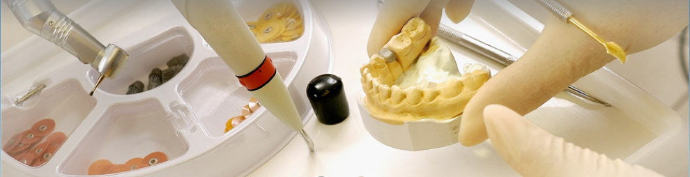
![]() We can start mounted studies models on :
We can start mounted studies models on :
- Examination records
- Pre-op photographs
- Radiographs
After the creating the mounted studies models, we start the diagnostic wax-up process.
In order to successfully work through the diagnostic process the following records are necessary:
- Well impressed models capturing all oral anatomy.
- Facebow
- Centric – Relation bite (CR)
- Protrusive bite
- Notes of clinical findings for any questionable teeth
- Patient’s expectations (whiter, straighter, longer, shorter teeth)
- Diagnostic Photos:
- Full face smile : should have only full face and shoulders in it
- Close-up smile: should be nose to chin
- Close-up at rest: lips slightly apart from nose to chin
- Close up high smile: this will show the relation between the upper lip and gingival contour
- Close-up profile: patient smiling – this will show the relation between upper incisal edge and lower lip
- Close-up , top to down, nose to chin- patient smiling- this will show the upper arch against the lower lip

